

June 5th - June 6th, 2019
Telč, Czech Republic
| Sascha Senck | |
| Upper Austria University of Applied Sciences Wels | |
 | Sascha Senck received his PhD degree at the University of Vienna (Austria) in 2012. He is currently working as research project manager at the Research Group Computed Tomography of the University of Applied Sciences Upper Austria (Campus Wels). He manages several research projects in the field of biomedical engineering and non-destructive testing. His research focuses on the three-dimensional characterization of bio-based materials and biological hard tissue using X-ray and phase contrast microcomputed tomography. |
X-ray microcomputed tomography of pathological bone samples in musculoskeletal research | |
Sascha Senck | |
| Early and accurate diagnosis of musculoskeletal disorders like arthrosis, osteoporosis, or diabetic foot syndrome can help reduce the incidence of fractures, infection-related complications, or severe amputation of limbs. Conventional radiography, X-ray computed tomography (CT), scintigraphy, magnetic resonance imaging, ultrasound, and positron emission tomography are currently the most important procedures for assessing complications associated with musculoskeletal disorders. However, none of these modalities is able to provide comprehensive information on pathological pathways, hence a multimodal approach should ideally be used for diagnosis, including the study of bone microstructure using X-ray microcomputed tomography (XCT). | |
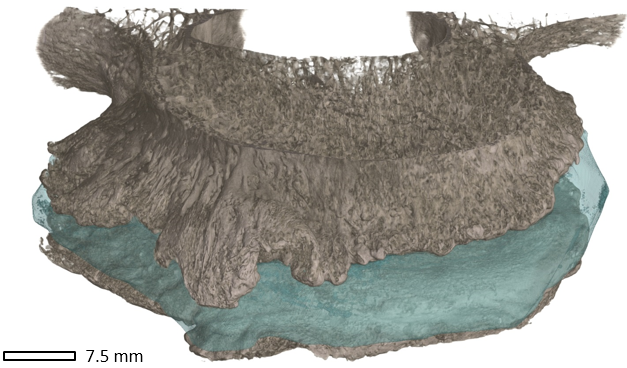 Figure 1: Endplates of lumbar vertebral bodies L2 and L3 from a human cadaver showing extensive osteophytes and a degenerated intervertebral disc (blue) |
For example, recent studies on the effects of type 2 diabetes mellitus on bone structure and strength use high-resolution peripheral quantitative CT (HR-pQCT). The resolution of second-generation HR-pQCT systems is high enough (isotropic voxel size of 61 µm) to represent the coarse trabecular bone structure and larger subchondral pores in vivo. However, for bone microstructure analyses at a higher spatial resolution, bone fragments can currently only be analyzed using XCT. Using the latest XCT system voxel sizes in the submicrometer range are possible. Numerous studies on the quantification of bone microstructure demonstrated the possibilities of three-dimensional visualization of the trabecular network and cortical bone using XCT and illustrate the benefits in (pre-)clinical research. Volume data serves to determine parameters of bone architecture such as number of trabeculae, mean trabecular thickness, and cortical thickness, as well as a basis for XCT-based biomechanical simulations. In this contribution we show several examples from musculoskeletal research including gonarthrosis (arthrosis of the knee), bone and cartilage degradation in osteoporosis of the spine (Fig. 1), and diabetic foot complications. We show that XCT is well suited to qualitatively and quantitatively characterize pathological bone and soft-tissue changes. Furthermore, we show examples using XCT data as starting point for biomechanical investigations using finite element analyses (FEA). |